Anatomy of the Eye
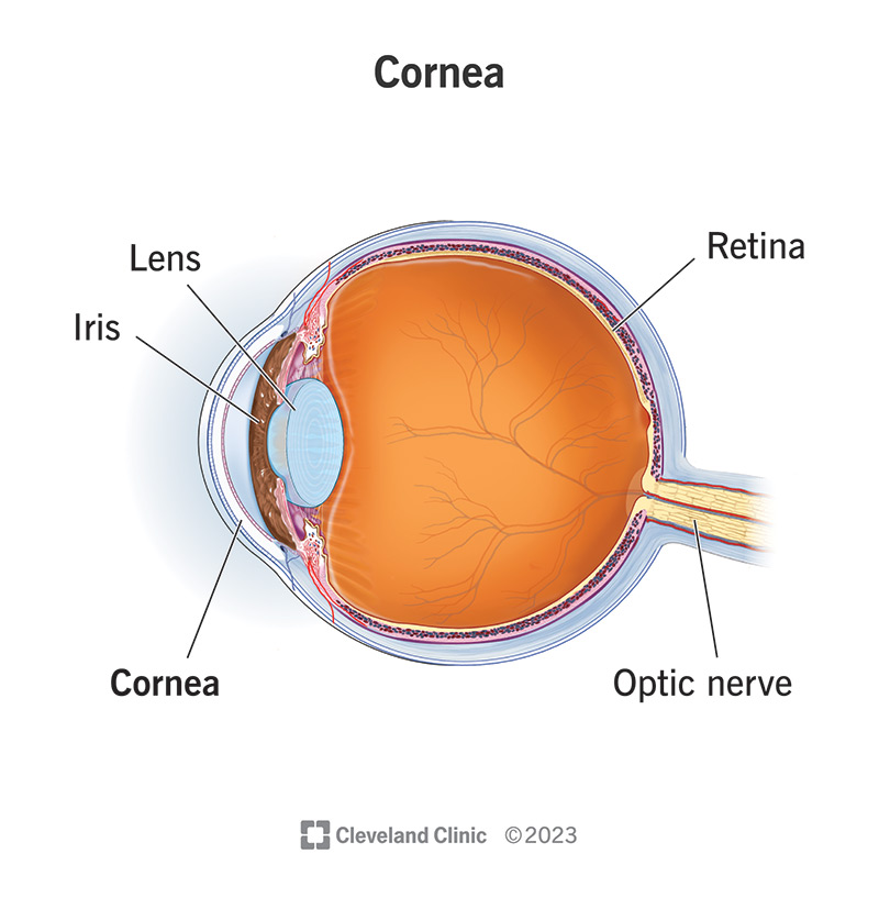
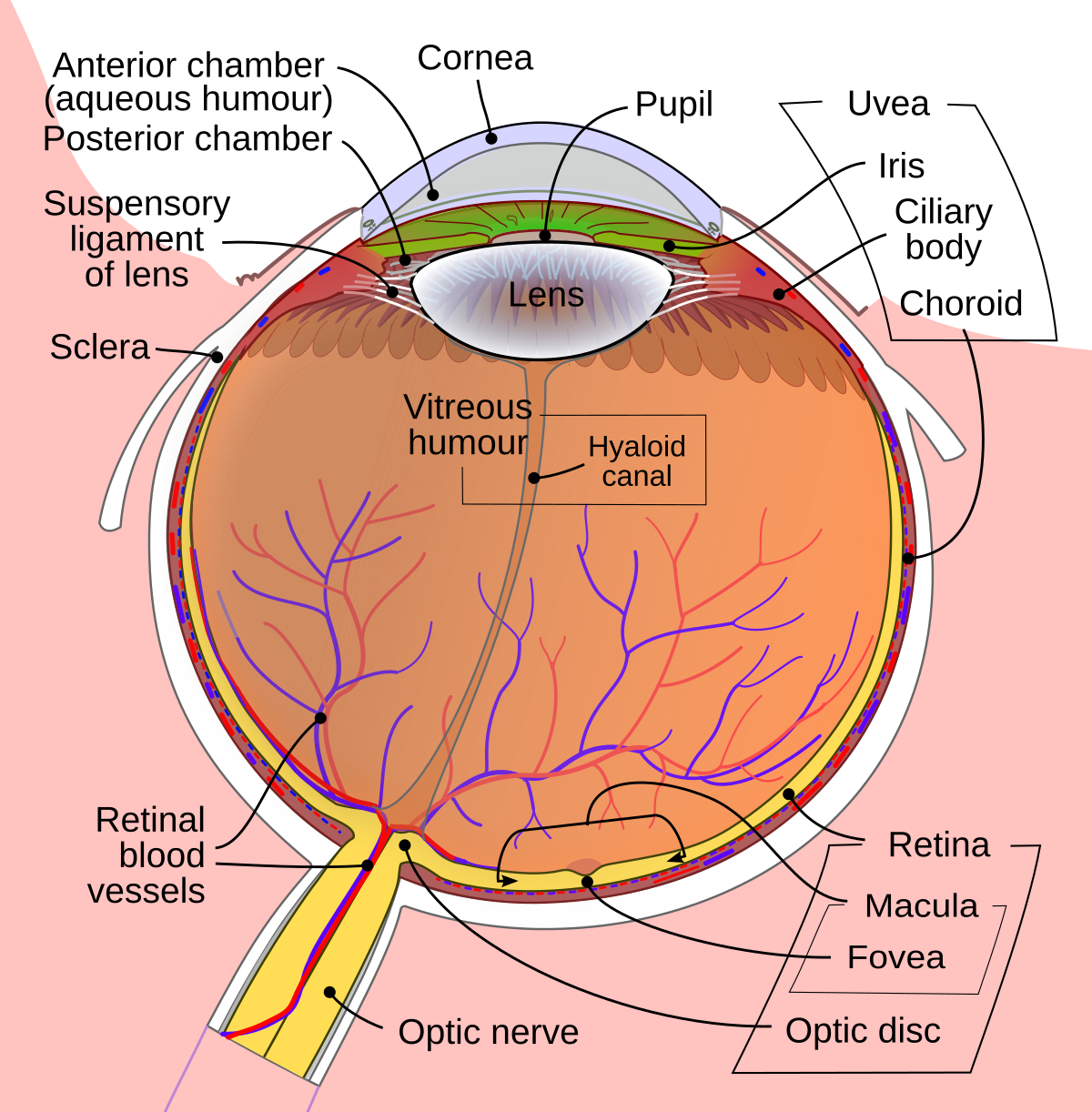
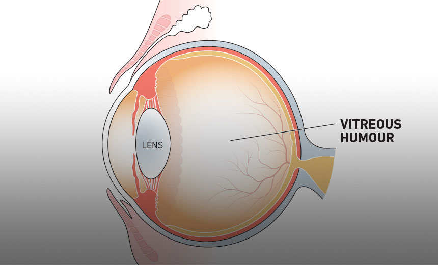
Illustrated Schematic of the Human Eye:
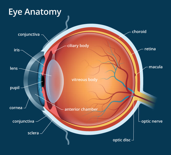
The human eye is a highly specialized organ that plays a critical role in visual perception, one of the primary sensory systems enabling interaction with the environment. As a complex optical system, the eye is designed to capture, process, and transmit light-derived information to the brain, where it is interpreted as visual images. Its intricate anatomy and physiology reflect a balance of structural precision and functional efficiency, ensuring adaptability to varying lighting conditions and focal demands. This text delves into the detailed anatomy of the human eye, exploring the roles of its individual components and their contributions to the overarching mechanism of vision.
Choroid
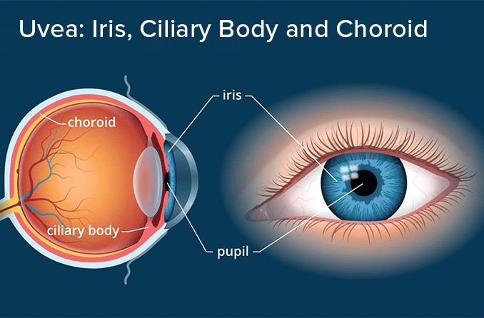
The choroid is a vascular layer located between the retina, the innermost light-sensitive tissue, and the scleroses, the outer white protective structure of the eye. This layer is rich in blood vessels, supplying oxygen and nutrients essential for retinal function while aiding in the removal of waste products.
Ciliary Body
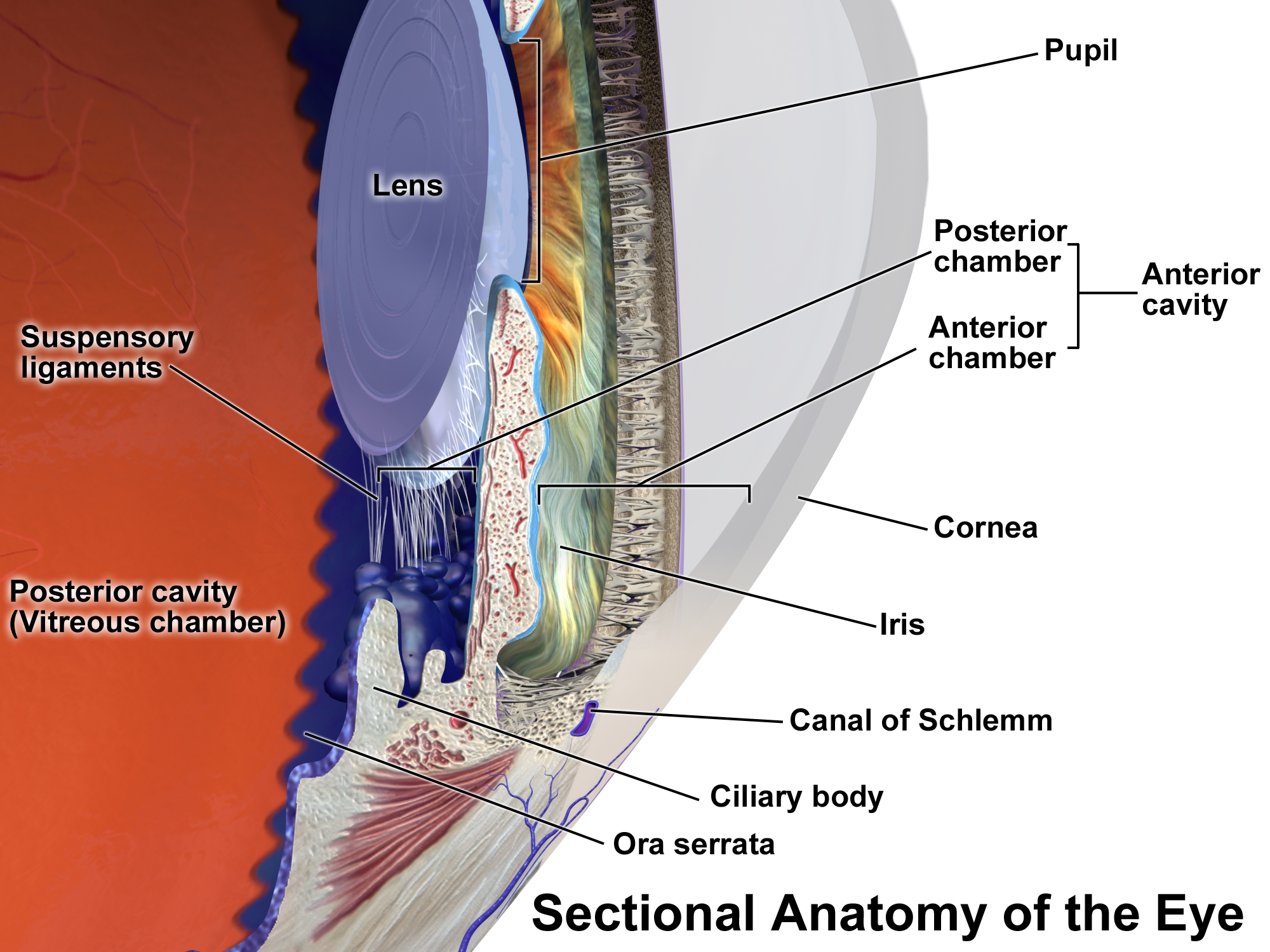
Positioned posterior to the iris, the ciliary body is a muscular structure responsible for accommodation, enabling the human eye to focus on objects at varying distances. It alters the shape of the lens and also produces aqueous humor, which maintains intraocular pressure and provides nutrients to a vascular structures of the eye.
Cornea
The cornea forms the human eye’s transparent anterior surface, serving as the primary refractive medium that focuses light onto the retina. Its curvature determines the sharpness of focus. Surgical procedures, such as laser-assisted in situ keratomileusis (LASIK), reshape the cornea to correct refractive errors.
Fovea
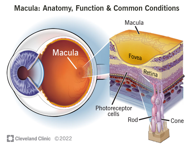
Centrally located within the macula, the fovea contains a dense concentration of cone photoreceptors, providing the highest visual acuity. It is critical for tasks requiring fine detail, such as reading and recognizing faces.
Iris
The iris is the pigmented, circular structure controlling the diameter of the pupil, thereby regulating the amount of light entering the human eye. In response to bright conditions, the iris contracts the pupil (miosis) to limit light intake, while under dim lighting, it dilates the pupil (mydriasis) to enhance light capture.
Lens
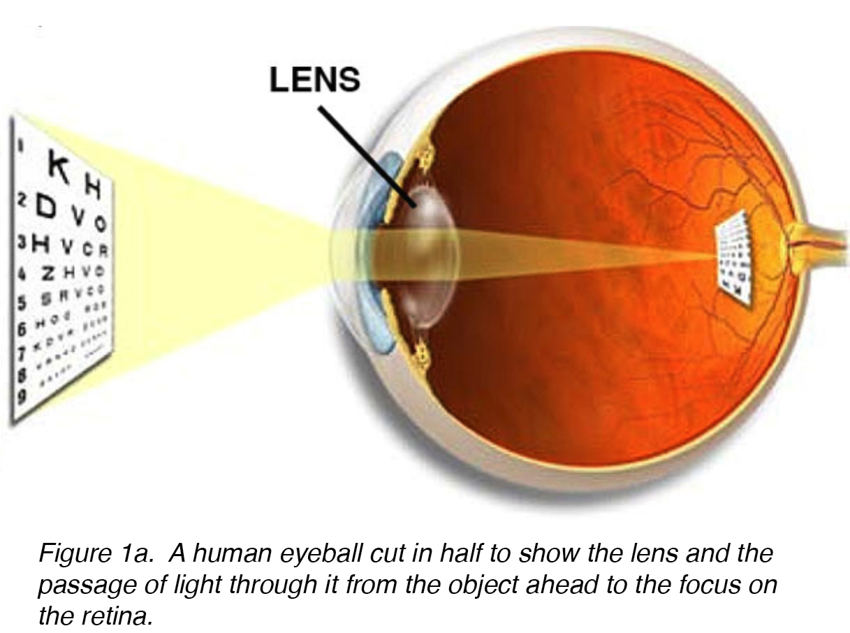
The crystalline lens is a biconvex, transparent structure responsible for fine-tuning the focus of light onto the retina. Aging and disease can lead to lens opacification, known as cataracts, which impair vision. Intraocular lenses are used to replace compromised lenses and restore clarity.
Macula
The macula is a specialized central region of the retina containing a high density of photoreceptors, particularly cones. It facilitates central vision, enabling the perception of fine details. Degeneration of the macula, commonly age-related macular degeneration (ARMD), can lead to significant visual impairment.
Optic Nerve
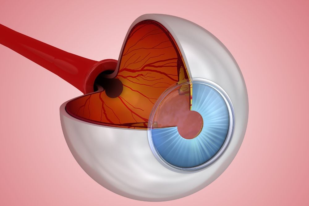
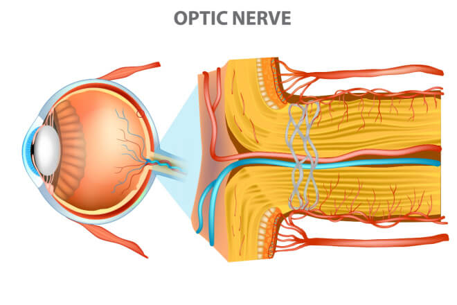
The optic nerve comprises over a million nerve fibers that transmit visual information from the retina to the brain’s visual cortex. This neural communication underpins the brain’s ability to process and interpret visual stimuli. Disorders such as glaucoma can result in optic nerve damage and vision loss.
Pupil
The pupil, the black circular aperture at the center of the iris, dynamically adjusts its size to modulate light entry. This function mimics a camera aperture, optimizing light levels for image clarity under varying lighting conditions.

Retina
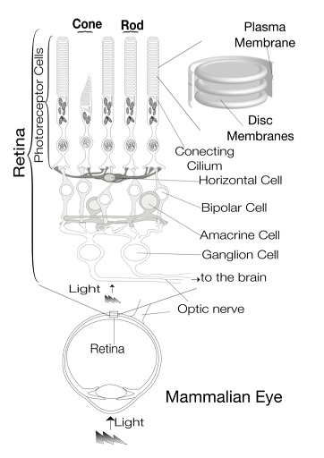
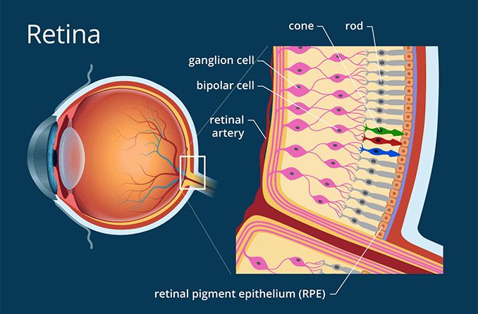
The retina, a multilayered neural tissue lining the inner posterior segment of the human eye, contains photoreceptor cells (rods and cones) that convert light into electrochemical signals. These signals are relayed via the optic nerve to the brain for image formation.
Sclera
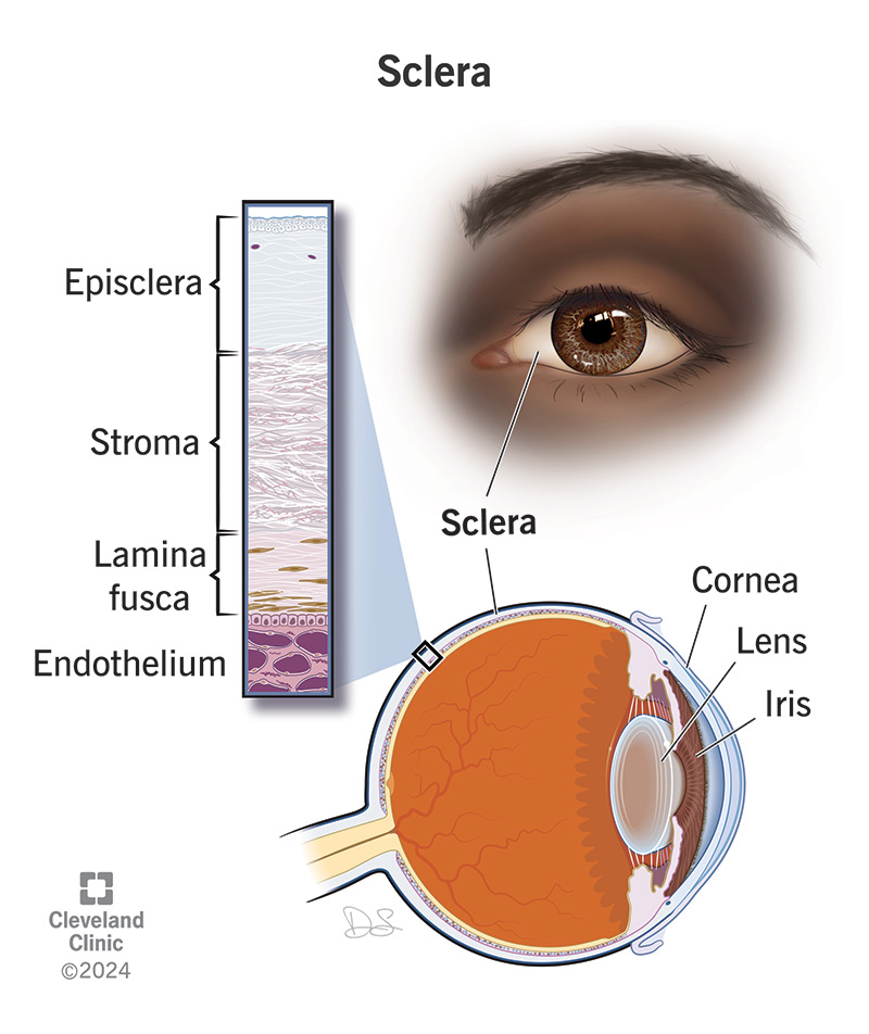
The sclera, a dense connective tissue layer, provides structural integrity and protection for intraocular contents. It also serves as an attachment site for extraocular muscles that facilitate eye movement.
Vitreous Humor
The vitreous humor is a clear, gel-like substance filling the eye’s vitreous chamber. It maintains ocular shape and ensures light transmission to the retina.
Mechanism of Vision

Vision involves a complex interplay between light, ocular structures, and neural pathways. Light enters the eye through the cornea, where initial refraction occurs. It then passes through the aqueous humor, pupil, and lens, which further adjusts focus before reaching the retina.
Within the retina, photoreceptor cells (rods for low light and cones for color vision) absorb the light, initiating a cascade of biochemical reactions that generate electrochemical signals. These signals are transmitted via the optic nerve to the brain’s visual processing centers, where they are interpreted as coherent images.
The eye’s functioning closely resembles a camera. The pupil operates as an adjustable aperture, modulating light entry, while the lens focuses light onto the retinal “film.” In cases of refractive errors, corrective measures such as spectacles, contact lenses, or surgical interventions restore optimal focus.

